
01fc1a8a7fcbfcc0e78fd82432ecd829.gif (2336×3018) Muscle diagram
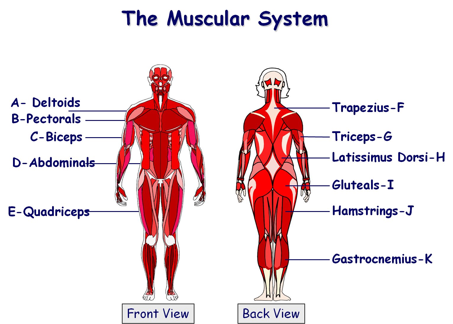
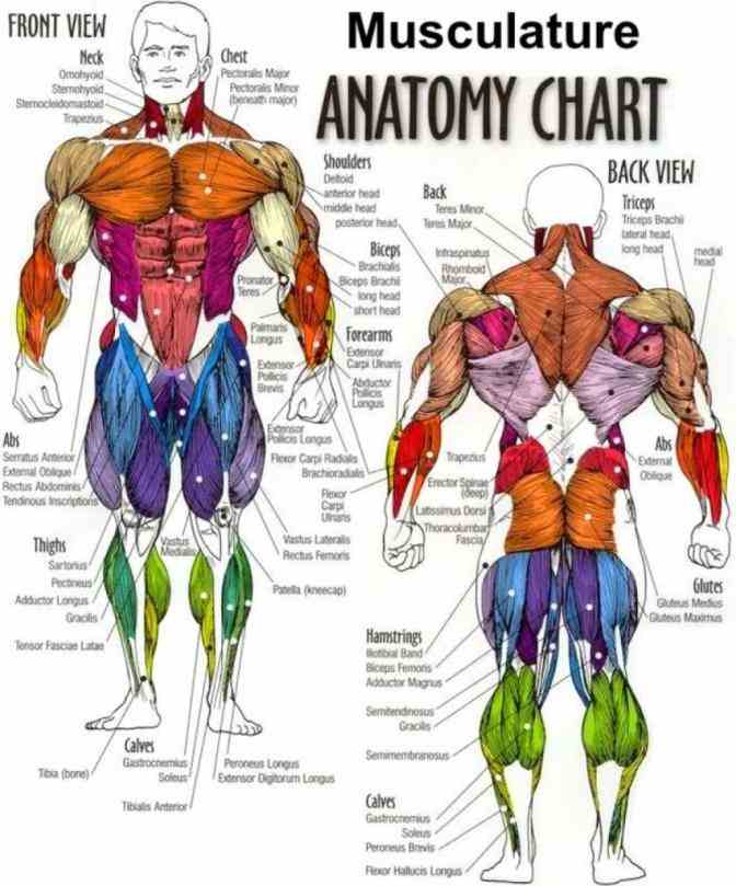
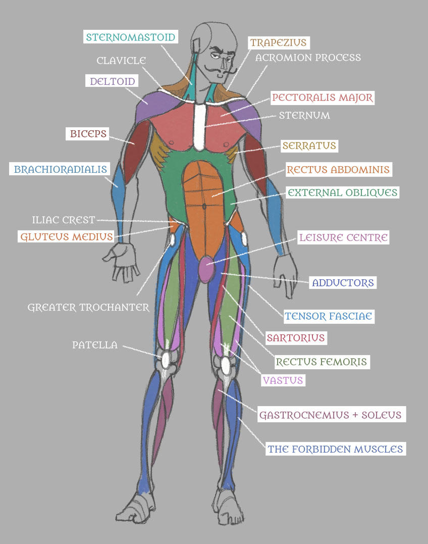
Here is the diagram of the human muscular system: Figure 5: Muscle chart showing the muscular system labeled where most muscles of the body are labeled in the form of a map of muscles.. 2- Surgical muscle-tendon transfer: it is important to understand and study the human muscle anatomy in many fields such as the surgical field. Since tendons.
Labeled Body Muscle Diagram Simple Labeled Muscle Diagram Human Body
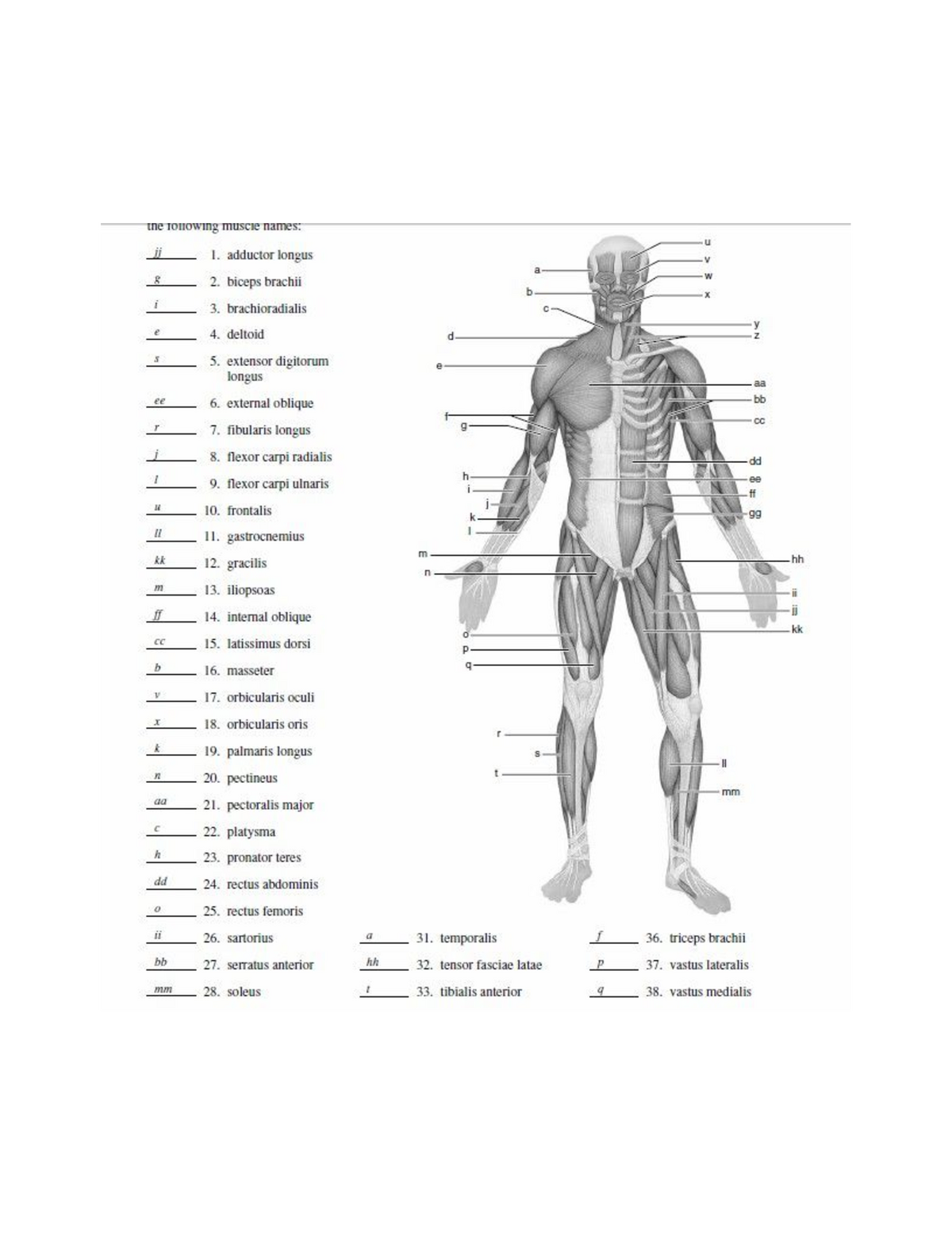
Human Anatomy, 6/e. Kent Van De Graaff, Weber State University. Muscular System. Labeling Exercises. Muscles-Anterior View 1 Muscles-Anterior View 2 Muscles- Anterior View 3 Leg Muscles-Anterior View 1 Leg Muscles-Anterior View 2 Muscles-Posterior View 1
Human Muscle Anatomy Diagram 433295 Vector Art at Vecteezy
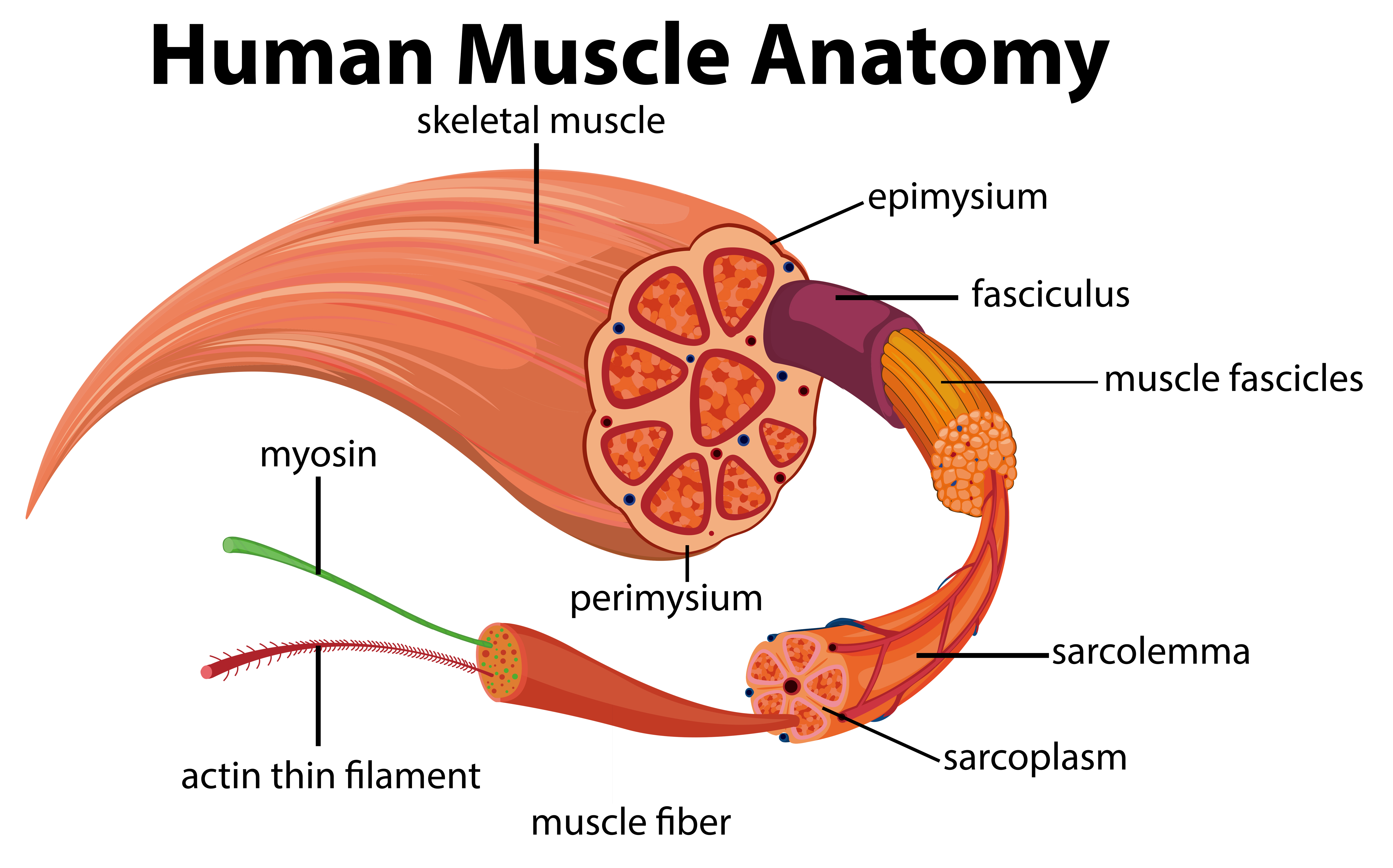
A typical myofiber is 2-3 centimeters ( 3/4-1 1/5 in) long and 0.05millimeters (1/500 inch) in diameter and is composed of narrower structures - myofibrils. These contain thick and thin myofilaments made up mainly of the proteins actin and myosin. Numerous capillaries keep the muscle supplied with the oxygen and glucose needed to fuel.

Muscles Labeled Front And Back / Muscle chart front view Do you even
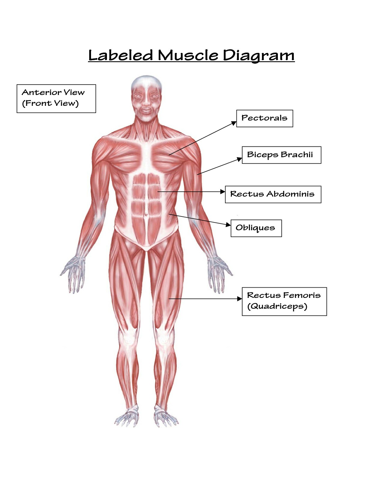
Human Anatomy - Front View of Muscles. Click on the labels below to find out more about your muscles. More human anatomy diagrams: back view of muscles, skeleton, organs, nervous system. Flex some.

FileMuscles anterior labeled.png Wikipedia
Muscles attach to bones directly or through tendons or aponeuroses. Skeletal muscles maintain posture, stabilize bones and joints, control internal movement, and generate heat. Skeletal muscle fibers are long, multinucleated cells. The membrane of the cell is the sarcolemma; the cytoplasm of the cell is the sarcoplasm.

FileMuscle posterior labeled.png Wikipedia
Muscular System / In these topics. Muscles. Brought to you by Merck & Co, Inc., Rahway, NJ, USA (known as MSD outside the US and Canada)—dedicated to using leading-edge science to save and improve lives around the world. Learn more about the MSD Manuals and our commitment to Global Medical Knowledge.
Human Body Mrs. Willis 7th Life Science
Each skeletal muscle is an organ that consists of various integrated tissues. These tissues include the skeletal muscle fibers, blood vessels, nerve fibers, and connective tissue. Each skeletal muscle has three layers of connective tissue (called "mysia") that enclose it and provide structure to the muscle as a whole, and also.

Back Muscles Diagram Labeled Labeled Muscle Diagram — UNTPIKAPPS We
The coracobrachialis is the smallest of the three muscles that attach to the coracoid process of the scapula. (The other two muscles that attach here are the pectoralis minor and the short head of the biceps brachii.) It is situated at the upper and medial part of the arm. It is supplied by the musculocutaneous nerve.

Muscle Diagram You Can Do More!
The muscular system is responsible for the movement of the human body. Attached to the bones of the skeletal system are about 700 named muscles that make up roughly half of a person's body weight. Each of these muscles is a discrete organ constructed of skeletal muscle tissue, blood vessels, tendons, and nerves.
Labeled Body Muscle Diagram
Muscle Anatomy. The interactive muscle anatomy diagram shown below outlines the major superficial (i.e. located immediately below the skin) muscles of the body. It should be noted that there are many more muscles in the body that are not addressed by this muscle anatomy diagram, however the muscles that are of primary interest from a fitness.

Muscle Diagram Most Important Muscles Of An Athletic Male Body Anterior
Anatomy and Physiology Nursing Test Banks. This nursing test bank includes questions about Anatomy and Physiology and its related concepts such as: structure and functions of the human body, nursing care management of patients with conditions related to the different body systems. Dive into the ultimate study guide for the muscular system.

Labeled Muscle Diagram Chart Free Download
Muscular. The primary job of muscles is to move the bones of the skeleton, but muscles also enable the heart to beat and constitute the walls of other vital hollow organs. Skeletal muscle: This.
Labelled Muscular System Front And Back Muscles of the Body Quiz
externus. outside. EXternal. internus. inside. INternal. Anatomists name the skeletal muscles according to a number of criteria, each of which describes the muscle in some way. These include naming the muscle after its shape, its size compared to other muscles in the area, its location in the body or the location of its attachments to the.
Simple Human Muscles Diagram Major Muscles Of The Human Body For Kids
Leg muscles (Musculi cruris) Anatomically, the leg is defined as the region of the lower limb below the knee. It consists of a posterior, anterior and lateral compartment. In accordance, the muscles of the leg are organized into three groups: Anterior (dorsiflexor) group, which contains the tibialis anterior, extensor digitorum longus.
Blank Muscle Diagram to Label ANP1106 uOttawa StuDocu
Inner hip & gluteal muscles. Anterior, medical and posterior thigh muscles. Anterior, lateral and posterior leg muscles. Dorsal and plantar foot muscles. This eBook contains high-quality illustrations and validated information about each muscle. It is available for free. Download free PDF (8.5MB) Get for free on iBooks.

Labeled Muscles In The Body Diagram Black And White Muscular System
Gastrocnemius (calf muscle): One of the large muscles of the leg, it connects to the heel. It flexes and extends the foot, ankle, and knee. Soleus: This muscle extends from the back of the knee to.